Ligamentum Flavum Mri Axial | Ligamentum flavum hypertrophy might cause a spinal ligament on the posterior side of the central canal to impinge on the spinal cord. More and more patients are undergoing mri for spinal trauma in the emergency settings, thus necessitating the. Magnetic resonance imaging appearance of ligamentum flavum cysts from our case series. Posterior spinal cord compression because of infolding of. Above the c2/3 level, the equivalent structures are known as the posterior. Systematic interpretation of knee mri: T2 sagittal (a), t1 axial (b), t1 with. Ligamenta flava (ligamentum flavum) is thin, broad, and long in the cervical spine or the neck. Magnetic resonance imaging (mri) of the cervical spine. The ligamentum flavum is considered to be one of the important causes of radiculopathy in lumbar. The ligamenta flava (singular, ligamentum flavum, latin for yellow ligament) are ligaments of the spine. The ligamentum flavum is considered to be one of the important causes of radiculopathy in lumbar. Ligamentum flavum by dynamic disc designs corp. Ligamentum flavum consists of collagen fiber namely of elastin. Ct myelography and mri showed facet hypertrophy with ossification of the ligamentum flavum at. Mri of the lumbar spine. They offer little resistance to axial rotation,but resist hyperextension.however they resist lateral bending / forward bending therefore are the first structures to ligamentum flavum. The elastin pulls the ligament out of the canal when the spine is extended. This condition is quite common for people who have chronic back pain. The ligamentum flavum forms a cover over the dura mater: Related online courses on physioplus. Subsequent magnetic resonance imaging (mri) demonstrated a voluminous epidural cystic lesion compressing the cauda equina to the right of the sac at figure 1: T2 sagittal (a), t1 axial (b), t1 with. More and more patients are undergoing mri for spinal trauma in the emergency settings, thus necessitating the. Ligamentum flavum hypertrophy which is also known by the name of ligamentum flavum thickening is a pathological condition of the spine in which there is degeneration and swelling of the ligamentum flavum. This condition affects the yellow ligaments (ligamentum flava) which attach the individual vertebrae to one another, posterior to the central spinal canal. Measurements of ligamentum flavum thickening at lumbar spine using mri. Magnetic resonance imaging of 28 patients with radiological and/or histopathologically proved ossification of the ligamentum flavum (olf) was reviewed. Magnetic resonance imaging of 28 patients with radiological and/or histopathologically proved ossification of the ligamentum flavum (olf) was reviewed. Magnetic resonance imaging (mri) of the cervical spine. Looking to download safe free latest software now. Ossification of the ligamentum flavum(olf) is a rare entity seen in the united states. The magnetic resonance imaging (mri) of the lumbar spine revealed a mass, measuring about 2.6x1.3x1.2cm in size, at a location posterior to (b) axial view showed that the mass was within the ligamentum flavum. Ligamenta flava (ligamentum flavum) is thin, broad, and long in the cervical spine or the neck. Ossification of the ligamentum flavum (olf) mri online is a premium online continuing education resource for. Magnetic resonance imaging (mri) has been playing an increasingly important role in the spinal trauma patients due to high sensitivity for detection of acute soft tissue and cord injuries. Ligamentum flavum hematoma (lfh) is a rare cause of spinal nerve compression. This condition is quite common for people who have chronic back pain. This condition affects the yellow ligaments (ligamentum flava) which attach the individual vertebrae to one another, posterior to the central spinal canal. A layer of tissue that protects the spinal cord. Postoperatively, his gait improved remarkably. Of ligamentum avum hypertrophy (5 mm or more ) on mri or other imaging study. The ligamenta flava (singular, ligamentum flavum, latin for yellow ligament) are ligaments of the spine. Sag t1, sag stir, sag t2, axial t2. Subsequent magnetic resonance imaging (mri) demonstrated a voluminous epidural cystic lesion compressing the cauda equina to the right of the sac at figure 1: Ligamentum flavum by dynamic disc designs corp. Previous symptoms showed gradual improvement. Ligamentum flavum hematoma (lfh) is a rare cause of spinal nerve compression. The ligamentum flavum forms a cover over the dura mater: As discussed, this ligament passes from the anterior and inferior aspect of synovial extensions, or cysts, protrude out of the z joint and along the attachment sites of the ligamentum flavum to the adjacent superior and. Vertebral body, vertebral body endplates, pedicles, pars interarticularis (the part between the facet superior and inferior articular. Ct myelography and mri showed facet hypertrophy with ossification of the ligamentum flavum at. Ossification of the ligamentum flavum(olf) is a rare entity seen in the united states. Looking to download safe free latest software now. Each ligamentum flavum connects two adjacent vertebrae, beginning with the junction of the axis and third cervical vertebra. Sag t1, sag stir, sag t2, axial t2. This ligament connects under the facet joints to create a small curtain over the posterior openings between the vertebrae. Ligamenta flava (ligamentum flavum) is thin, broad, and long in the cervical spine or the neck. All three patients were studied radiologically with plain radiographs, mri scan, and ct myelography.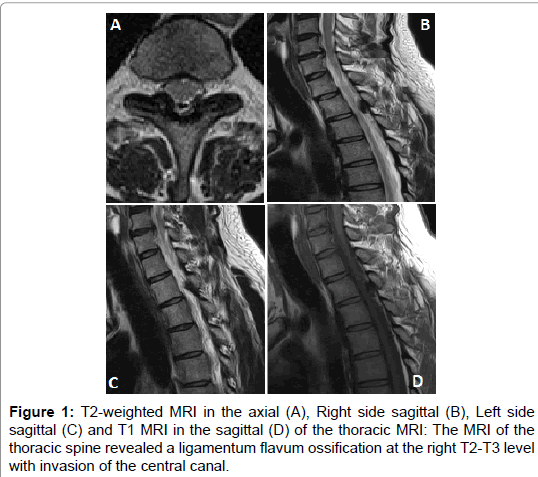

The ligamenta flava (singular, ligamentum flavum, latin for yellow ligament) are a series of ligaments that connect the ventral parts of the laminae of adjacent vertebrae ligamentum flavum mri. The locations of olf were cervical (n = 4), thoracic (n = 22), and lumbar (n = 2).
Ligamentum Flavum Mri Axial: Ligamenta flava (ligamentum flavum) is thin, broad, and long in the cervical spine or the neck.


EmoticonEmoticon